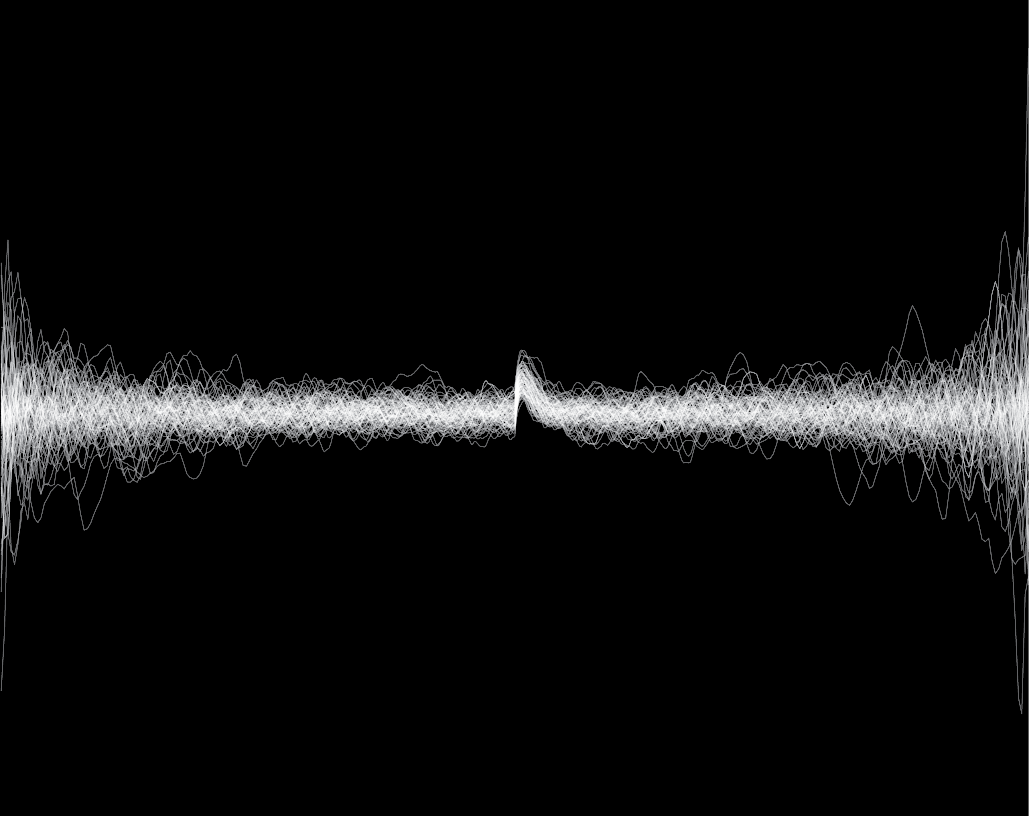

The H2A.Z histone variant, a genome-wide hallmark of permissive chromatin, is enriched near transcription start sites in all eukaryotes. H2A.Z is deposited by the SWR1 chromatin remodeler and evicted by unclear mechanisms. We tracked H2A.Z in living yeast at single-molecule resolution, and found that H2A.Z eviction is dependent on RNA Polymerase II (Pol II) and the Kin28/Cdk7 kinase, which phosphorylates Serine 5 of heptapeptide repeats on the carboxy-terminal domain of the largest Pol II subunit Rpb1. These findings link H2A.Z eviction to transcription initiation, promoter escape and early elongation activities of Pol II. Because passage of Pol II through +1 nucleosomes genome-wide would obligate H2A.Z turnover, we propose that global transcription of noncoding RNAs prior to premature termination, in addition to transcription of mRNAs, are responsible for eviction of H2A.Z. Such usage of yeast Pol II suggests a general mechanism coupling eukaryotic transcription to erasure of the H2A.Z epigenetic signal.
Live-cell single particle imaging reveals the role of RNA polymerase II in histone H2A.Z eviction
Ranjan A, Nguyen VQ, Liu S, Wisniewski J, Kim JM, Tang X, Mizuguchi G, Elalaoui E, Nickels TJ, Jou V, English BP, Zheng Q, Luk E, Lavis LD, Lionnet T, Wu C. (2020)

The spatial organization of the genome inside the nucleus is known to impact gene expression, for instance through physical contacts between the promoter of a gene and a regulatory DNA sequence localized far away along the chromosome. How DNA folding impacts the expression of key cancer driving genes is unclear. Here, we characterize the interplay between DNA organization and gene expression in T cell acute lymphoblastic leukemia (T-ALL) cells, demonstrating a change in physical organization linked with cancer cells at the Myc locus, correlated with expression of the oncogene.
Dynamic 3D chromosomal landscapes in acute leukemia
Kloetgen K*, Thandapani P*, Ntziachristos P*, Ghebrechristos Y, Nomikou S, Lazaris C, Chen X, Hu H, Bakogianni S, Wang J, Fu Y, Boccalatte F, Zhong H, Paietta E, Trimarchi T, Zhu Y, van Vlierberghe P, Inghirami G, Lionnet T, Aifantis I and Tsirigos A. (2019) BiorXiv | Nature Genetics

Embryos initially contain parental mRNA and do not transcribe their own genes. They remain silent until the Zygotic Genome Activation, when the first genes are transcribed. How this happens, and how chromatin changes to favor the emergence of transcription remains unclear. This work demonstrates how fluorescently labeled antibody fragments (Fabs) can be used to track the changes in chromatin modifications that pave the way for the onset of transcription.
Histone H3K27 acetylation precedes active transcription during zebrafish zygotic genome activation as revealed by live-cell analysis
Sato Y, Hilbert L, Oda H, Wan Y, Heddleston JM, Chew TL, Zaburdaev V, Keller P, Lionnet T, Vastenhouw N, Kimura H.
(2019) Development 146, dev179127 | BiorXiv

mRNA quantification using single-molecule FISH in Drosophila embryos.
Trcek T, Lionnet T, Shroff H, Lehmann R.
(2017) Nature Protocols 12(7):1326-1348.

Quantitative mRNA Imaging Throughout the Entire Drosophila Brain
Long X*, Colonell J, Wong AM, Singer RH, Lionnet T*, (2017) Nature Methods 14(7):703-706; Bioarxiv
*Corresponding author

Bright photoactivatable fluorophores for single-molecule Imaging.
Grimm JB, English BP, Choi H, Muthusamy AK, Mehl BP, Dong P, Brown TA, Lippincott-Schwartz J, Liu Z, Lionnet T*, Lavis LD* (2016) Nature Methods doi:10.1038/nmeth.4034 Bioarxiv PMC
*Corresponding author
Real-time Quantification of single RNA translation dynamics in living cells.
Morisaki T, Lyon K, DeLuca KF, DeLuca JG, English BP, Zhang Z, Lavis LD, Grimm JB, Viswanathan S, Looger LL, Lionnet T, Stasevich T. (2016) Science Jun 17; 352 (6292): 1425-9

RNA Polymerase II cluster dynamics predicts mRNA ouput in living cells.
Cho WK, Jayanth N, English BP, Inoue T, Andrews JO, Conway W, Grimm JB, Spille JH, Lavis LD, Lionnet T*, Cisse II* (2016) eLife PMC
*Corresponding author

Multifocus microscopy with precise color multi-phase diffractive optics applied in functional neuronal imaging.
Abrahamsson S, Ilic R, Wisniewski J, Mehl B, Yu L, Chen L, Davanco M, Oudjedi L, Fiche J-B, Hajj B, Jin X, Pulupa J, Cho C, Mir M, El Beheiry M, Darzacq X, Nollmann M, Dahan M, Wu C, Lionnet T, Liddle AJ, Bargmann CI (2016) Biomedical Optics Express 7 (3) 855 PMC

Mapping translation ‘hot spots’ in live cells by tracking single molecules of mRNA and ribosomes.
Katz ZB, English BP, Lionnet T, Yoon YJ, Monnier N, Ovryn B, Bathe M, Singer RH. (2016) eLife 10415 PMC

CASFISH : CRISPR/Cas9-mediated in situ labeling of genomic loci in fixed cells.
Deng W, Shi X, Tjian R, Lionnet T, Singer RH. (2015) Proc. Natl. Acad. Sci., 112 (38), 11870-11875 PMC

Drosophila germ granules are structured and contain homotypic mRNA clusters.
Treck T, Grosch M, York A, Shroff H, Lionnet T, Lehman R. (2015) Nature Communications, 6:7962 PMC

Cellular Levels of Signaling Factors Are Sensed by β-actin Alleles to Modulate Transcriptional Pulse Intensity.
Kalo A, Kanter I, Shraga A, Sheinberger J, Tzemach H, Kinor N, Singer RH, Lionnet T, Shav-Tal Y. (2015) Cell Reports 11(3) 419-32 PMC

An RNA biosensor for imaging the first round of translation from single cells to living animals.
Halstead JM*, Lionnet T*, Wilbertz JH*, Wippich F*, Ephrussi A, Singer RH, Chao JA. (2015) Science 347(6228) 1367-671 PMC
*Equal Contributions

A general method to improve fluorophores for live-cell and single-molecule microscopy.
Grimm JB, English BP, Chen J, Slaughter JP, Zhang Z, Revyakin A, Patel R, Macklin JJ, Normanno D, Singer RH, Lionnet T*, Lavis LD.* (2015) Nature Methods 12(3) 244-50 PMC
*Corresponding author

Single-molecule dynamics of enhanceosome assembly in embryonic stem cells.
Chen J, Zhang Z, Li L, Chen BC, Revyakin A, Hajj B, Legant W, Dahan M, Lionnet T, Betzig E, Tjian R, Liu Z. (2014) Cell 156 (6) 1274-85 PMC

Colocalization of different influenza viral RNA segments in the cytoplasm before viral budding as shown by single-molecule sensitivity FISH analysis.
Chou YY, Heaton NS, Gao Q, Palese P, Singer R, Lionnet T.* (2013) PLoS Pathogens, 9 (5) e1003358 PMC
- Corresponding Author

Transcription goes digital.
Lionnet T, Singer RH (2012). Embo Reports, 13(4):313-21 PMC

Spatial arrangement of conserved recognition elements identifies RNA regulatory networks.
Patel VL, Mitra S, Harris R, Buxbaum AR, Lionnet T, Girvin M, Levy M, Almo SC, Brenowitz M, Singer RH, Chao JA. (2012) Genes & Development, 26 (1) 43-53; PMC

A transgenic mouse for in vivo detection of endogenous labeled mRNA.
Lionnet T, Czaplinski K, Darzacq X, Shav-Tal Y, Wells AL, Chao JA, Park HY, de Turris V, Lopez-Jones M, Singer RH (2011). Nature Methods, 8(2) 165-70 PMC

Transcription of functionally related genes is not coordinated.
Gandhi SJ, Zenklusen D, Lionnet T, Singer RH (2011). Nature Structural & Molecular Biology, 18 (1) 27-34 PMC

Real-time observation of bacteriophage T4 gp41 helicase reveals an unwinding mechanism.
Lionnet T, Spiering MM, Benkovic SJ, Bensimon D, Croquette V (2007). Proc. Natl. Acad. Sci. 104 (50): 19790-5 PMC

Sequence dependent twist-stretch coupling in DNA.
Lionnet T, Lankas F (2007). Biophys. J. 92 (4): L30-32 PMC

Wringing out DNA.
Lionnet T, Joubaud S, Lavery R, Bensimon D, Croquette V (2006). Phys. Rev. Lett., 96 (17): 178102 PMC

Single-molecule assay reveals strand switching and enhanced processivity of UvrD.
Dessinges MN, Lionnet T, Xi XG, Bensimon D, Croquette V (2004). Proc. Natl. Acad. Sci. 101 (17): 6439-44 PMC